
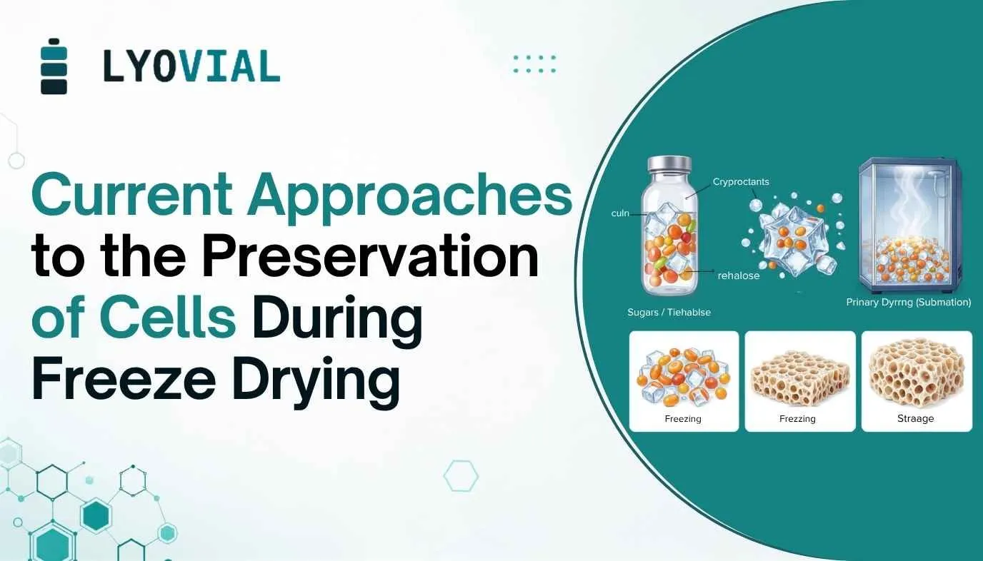
Executive Summary
Therapeutic and research cells — stem cells, blood components, spermatozoa, and microorganisms — are almost universally preserved today by cryopreservation at or below −130 °C. This delivers reliable viability but imposes a persistent cold chain, with substantial transport and storage costs and the risk of transient warming events. (Freeze-)drying offers an alternative: converting cells into a stable, low-moisture solid that can, in principle, be stored and shipped at ambient or refrigerated temperatures and reconstituted rapidly on demand.
This white paper synthesizes the current scientific understanding of cell damage during freezing, drying, and rehydration, and surveys the excipient classes and process strategies used to counteract that damage. Trehalose remains the single most effective and most widely used stabilizer, particularly when it can be delivered to both sides of the plasma membrane; sugars, macromolecules, polyols, antioxidants, and chelating agents each contribute complementary protection, but no universal “stabilization cocktail” yet exists. Success is highly cell-type dependent: microorganisms and spermatozoa already tolerate drying well enough for practical use, whereas complex, terminally differentiated eukaryotic cells remain the most difficult targets, with proliferative recovery so far reported almost exclusively for stem and progenitor cell populations.
The paper closes with a review of process-level levers — freezing rate, residual moisture content, rehydration protocol, and initial cell density — and with a short outlook on recent (2022–2026) developments, including improved sugar/protein co-formulations for cell-derived secretomes and continued refinement of intracellular trehalose delivery.
Table of Contents
1. Introduction
2. Mechanisms of Cellular Damage During (Freeze-)Drying
2.1 Freezing
2.2 Primary and Secondary Drying
2.3 Rehydration
2.4 Alternative Drying Methods
3. Protective Excipients
3.1 Sugars
3.2 Macromolecules
3.3 Polyols
3.4 Antioxidants
3.5 Chelating Agents
3.6 Excipient Toxicity
4. Intracellular Excipient Loading Technologies
5. Process Parameters
5.1 Freezing Rate
5.2 Residual Moisture Content
5.3 Rehydration Conditions
5.4 Cell Density
6. Current Approaches by Cell Type
6.1 Microorganisms
6.2 Red Blood Cells and Platelets
6.3 Spermatozoa
6.4 Eukaryotic Cells
7. Recent Developments (2022–2026)
8. Conclusions and Outlook
References
1. Introduction
Therapeutic cells are used across an expanding range of applications, including stem-cell transfer, wound healing, reproductive medicine, and transfusion support. These applications, together with the growth of regenerative medicine, require a constant and reliable supply of active cells. Ensuring that supply currently depends almost entirely on cryopreservation at temperatures below −130 °C, where all water is solidified and diffusion-limited chemistry effectively stops. Cryopreservation is robust, but it is also expensive to sustain: liquid-nitrogen storage, dry-ice or cryogenic shipping, and continuous monitoring all add cost, and any lapse — a transient warming event, a shipping delay, a freezer failure — can destroy an entire batch of cells.
Drying, and particularly freeze-drying (lyophilization), is an attractive alternative. A well-dried product can be stable at refrigerated or even ambient temperature, is far lighter and cheaper to transport, and — because freeze-drying leaves behind a defined, porous cake structure — reconstitutes quickly and controllably. The obstacle is biological rather than engineering: freezing, primary and secondary drying, and reconstitution each present a distinct opportunity for lethal cell injury, and unlike small-molecule drugs or even many proteins, a living cell has to survive the complete removal and subsequent replacement of its aqueous environment without losing membrane integrity, metabolic competence, or genomic integrity.
This white paper summarizes where the field currently stands: what actually damages cells during each phase of drying, which excipients and process choices mitigate that damage, and how successful current approaches are across microorganisms, blood components, spermatozoa, and eukaryotic cells. Because achieving long-term dry-state stability requires lengthy storage studies that many published protocols have not yet completed, most of the results discussed below describe cell condition immediately after drying rather than after extended storage; where storage data exist, that is noted explicitly.
2. Mechanisms of Cellular Damage During (Freeze-)Drying
2.1 Freezing
Freeze-drying begins with freezing, and the kinetics of ice formation determine much of what follows. As an aqueous cell suspension cools, ice nucleates first in the extracellular medium. The plasma membrane initially blocks ice from propagating into the cytoplasm, so intracellular water remains liquid and supercooled while extracellular solutes become progressively concentrated by the growing ice front. This creates a hypertonic extracellular environment; the cell responds by losing water osmotically and shrinking, a process often called “freeze-dehydration.” Within the resulting concentrated, crowded cytoplasm, proteins and organelles are packed closely enough that unwanted interactions and chemical degradation become more likely.
The cooling rate governs which failure mode dominates. At high cooling rates, water has insufficient time to leave the cell osmotically, so a larger fraction remains intracellular and eventually crystallizes; the resulting intracellular ice, and particularly its sharp crystal structure, can physically rupture organelles and membranes. At low cooling rates, cells have more time to dehydrate osmotically, largely avoiding intracellular ice but suffering more pronounced shrinkage and prolonged exposure to a concentrated, potentially toxic extracellular brine. An intermediate, optimized cooling rate — balancing intracellular ice formation against osmotic injury — is therefore usually the target, though the literature shows no single freezing protocol that is optimal across all cell types.
2.2 Primary and Secondary Drying
Once cells are frozen, vacuum is applied to sublime the frozen water (primary drying), followed by desorption of unfrozen, bound water at elevated shelf temperature (secondary drying). This step is especially punishing for the plasma membrane. In the hydrated state, water molecules hydrogen-bond to the polar head groups of membrane phospholipids, keeping them spaced apart; the membrane is fluid and has a phase-transition temperature (Tm) below physiological temperature. As drying removes this hydration shell, the head groups pack more closely, van der Waals interactions between adjacent lipids increase, and Tm rises — for example, from roughly −7 °C in the hydrated state to about 57 °C once dry for a model phospholipid (POPC). At room temperature, the dried membrane is consequently forced into a rigid, gel-like phase. This transition occurs unevenly across the membrane and is frequently accompanied by leakage of intracellular contents and, in some cases, local membrane fusion. The same loss of hydrogen-bonded water also affects intracellular proteins and structures, forcing previously separated molecules into contact and promoting aggregation.
2.3 Rehydration
Reconstitution is not formally part of the drying cycle, but it can complete the injury of cells that survived freezing and drying only marginally. As rehydration medium is added, excipients dissolve and the cell is briefly exposed to a strongly osmotic, non-equilibrium environment before water fully penetrates the intracellular space. At the membrane level, added water reverses the process described above: phospholipids regain their hydration shell and the membrane reverts from the gel-like to the liquid-crystalline phase. If this “reversed” phase transition occurs while excess free water is already present, it is again associated with leakage of intracellular material. Consequently, both the rate and temperature of rehydration, and the osmolality of the rehydration medium, materially affect final recovery.
2.4 Alternative Drying Methods
Freeze-drying is not the only desiccation route studied. Air-drying (evaporative drying under an air stream or in a desiccant chamber), vacuum-drying (evaporative drying under modest vacuum, ~70 Torr), and spin-drying (which promotes a more even residual-moisture distribution and faster drying) have all been used. These methods avoid freezing injury altogether and require far less specialized equipment, but they generally cannot reach the very low residual moisture contents achievable by lyophilization, and they expose cells to an osmotically active, concentrating solution for longer, at room temperature, where microbiological and oxidative reactions are not kinetically arrested. As a result, air- and vacuum-dried products are typically reported at higher residual moisture (often expressed as g H₂O/g dry weight rather than percent) and are frequently rehydrated and assayed immediately, rather than being stored as a genuinely dry, shelf-stable product.
3. Protective Excipients
Because freezing and drying are mechanistically distinct stresses, excipients are broadly divided into cryoprotectants (which protect chiefly during freezing/thawing) and lyoprotectants (which protect during both freezing and the drying step itself). The excipient classes used to stabilize cells during (freeze-)drying are summarized below and in Table 1.
3.1 Sugars
Trehalose is the dominant excipient in this field. It is a non-reducing disaccharide, found at high concentration in many naturally desiccation-tolerant (anhydrobiotic) organisms, and it protects dry membranes through two complementary mechanisms. First, under the “water-replacement hypothesis,” its small size and multiple hydroxyl groups let it hydrogen-bond directly to phospholipid head groups in place of water, keeping the head groups spaced apart and depressing Tm by roughly 10 °C relative to the fully hydrated membrane and by as much as 80 °C in the fully dry state — enough, in many cases, to keep the membrane in the liquid-crystalline phase throughout drying and rehydration. Second, trehalose vitrifies readily, forming a highly viscous amorphous glass as water is removed; this glass mechanically restricts molecular movement, suppressing membrane fusion and further leakage.
Trehalose's relatively high glass-transition temperature (Tg), compared with other sugars, is generally credited for its superior performance, and amorphous trehalose can also sequester small amounts of adsorbed water as a crystalline dihydrate without disturbing the surrounding amorphous phase, making trehalose-stabilized products comparatively tolerant of suboptimal storage humidity. Because trehalose is expensive at industrial scale, other sugars have also been evaluated: among disaccharides, sucrose gives results approaching trehalose in some systems (though it typically requires higher concentrations, given its weaker membrane interaction and greater tendency to crystallize), while lactose, maltose, and various mono- and trisaccharides have generally underperformed disaccharides. Critically, trehalose (and other small sugars) act mainly extracellularly unless deliberately introduced into the cytoplasm, because they do not readily cross intact plasma membranes — a limitation that has driven the intracellular loading technologies discussed in Section 4.
3.2 Macromolecules
Polymers such as hydroxyethyl starch (HES), dextran, polyvinylpyrrolidone (PVP), and serum albumins cannot cross the plasma membrane and therefore contribute nothing to intracellular stabilization or to depression of Tm. Their value lies instead in the physical properties of the dried matrix: because Tg generally rises with molecular weight, macromolecules confer a high Tg', permitting faster, more aggressive freeze-drying cycles, and a correspondingly high Tg in the final product, supporting storage at higher temperatures. Several polysaccharides also raise formulation viscosity, which independently slows water efflux and limits osmotic stress. The consistent finding across the literature is that macromolecules must be combined with a sugar; used alone, they do not adequately stabilize membranes. HES in particular is used extensively — combined with glucose to freeze-dry red blood cells, and combined with albumin and intracellular trehalose to freeze-dry hematopoietic stem and progenitor cells — while dextran and PVP have been used successfully with mononuclear cells and mesenchymal stem cells.
3.3 Polyols
Glycerol is the classic cryoprotectant: a small, water-miscible trihydric alcohol that forms hydrogen bonds readily, can cross the plasma membrane by facilitated or passive diffusion, and thereby buffers intracellular osmotic pressure during freezing. Its cryoprotective performance, however, does not translate well to drying. Glycerol has a very low Tg (−93 °C), depresses the Tg of any mixture it is added to, and in several studies has actively inhibited formation of a proper dry, freeze-dried cake. Multiple groups have failed to observe a protective effect of glycerol specifically during the drying step, even where it clearly helped during freeze/thaw cryopreservation — supporting the conclusion that glycerol is a good cryoprotectant but a poor lyoprotectant, and that its use in a drying formulation should be minimized and paired with an excipient that offsets its Tg-depressing effect.
Sorbitol and mannitol differ only in the stereochemistry of one hydroxyl group but behave quite differently on drying: mannitol tends to crystallize on warming above Tg', and the resulting crystals can physically rupture membranes and organelles, so mannitol must be kept in its metastable amorphous form to be useful. Both sugar alcohols are generally less effective membrane stabilizers than the disaccharides, though sorbitol has shown some benefit, particularly for long-term storage stability of dried lactic acid bacteria. Adonitol (ribitol) is a less common polyol that produced very high survival (up to 100%) across several lactic acid bacteria strains in early work, apparently because — unlike sorbitol or mannitol — it is not metabolized by these organisms and so is not depleted as an intracellular solute.
3.4 Antioxidants
Osmotic and cold stress during freezing, and desiccation stress during drying, both promote formation of reactive oxygen species (ROS), which can drive lipid peroxidation, protein oxidation, and DNA strand breaks. Antioxidants are used extensively in cryopreservation of reproductive cells but remain only lightly explored for (freeze-)drying more broadly. Arbutin, a plant-derived hydroquinone glucoside, improved viability and preserved osteogenic differentiation capacity of vacuum-dried mesenchymal stem cells, apparently partly through ROS inhibition and partly by inducing the heat-shock protein HSP70. (−)-Epigallocatechingallate (EGCG), a green-tea polyphenol, achieved up to 91% survival of freeze-dried mononuclear cells when combined with trehalose, though its anticarcinogenic and antiviral activity also inhibited proliferation after reconstitution unless excess EGCG was removed. In freeze-dried bovine sperm, the iron chelator Desferal® improved membrane integrity while the antioxidant enzyme catalase did not, suggesting that hydroxyl-radical formation via free iron, rather than hydrogen peroxide, is the dominant oxidative threat in that system.
3.5 Chelating Agents
In spermatozoa, membrane damage during drying disturbs intracellular ionic homeostasis and releases Ca²⁺/Mg²⁺-activated endonucleases that attack DNA. EDTA and EGTA, both of which complex these divalent cations, are now established excipients for freeze-drying sperm; EGTA is structurally similar to EDTA but less toxic and has a higher affinity for Ca²⁺, and low-dose EDTA (1 mM) has been shown to match the effect of much higher EGTA concentrations (50 mM), while high-dose EDTA (50 mM) becomes counterproductive. To date, chelating agents have been studied almost exclusively in the context of spermatozoa preservation; their applicability to other cell types has not been systematically reported.
3.6 Excipient Toxicity
Every protective excipient carries a toxicity trade-off that must be balanced against its stabilizing concentration. Small, membrane-impermeant sugars such as trehalose can themselves induce osmotic/hypertonic stress at high concentration — upconcentration during ice formation compounds this further — and elevated trehalose has been linked to increased hemolysis in red blood cells. EGCG's protective and toxic effects share a single mechanism (membrane interaction), so they cannot be fully separated. As a general rule drawn from the cryoprotectant literature, excipients that bind fewer water molecules while still supporting vitrification and suppressing water–water interactions tend to be less toxic; concentration optimization, not simply excipient selection, is therefore central to formulation development.
Table 1. Overview of excipient classes used to stabilize cells during (freeze-)drying
| Class | Representative agents | Primary mode of action | Key limitation |
| Sugars | Trehalose, sucrose, lactose, glucose, raffinose | Water replacement at phospholipid head groups; depression of the membrane phase-transition temperature (Tm); vitrification | Poor membrane permeability; hypertonic stress at high concentration; cost (trehalose) |
| Macromolecules | Hydroxyethyl starch (HES), dextran, PVP, BSA/HSA | High glass-transition temperature (Tg); viscosity that limits water efflux; amorphous-glass formation | Cannot cross membranes; ineffective alone, must pair with a sugar |
| Polyols | Glycerol, sorbitol, mannitol, adonitol | Hydrogen-bond formation; membrane permeation (glycerol); osmotic buffering | Low Tg depresses cake Tg (glycerol); mannitol crystallizes and ruptures membranes |
| Antioxidants | EGCG, arbutin, catalase | Scavenging of reactive oxygen species (ROS); some membrane interaction | Narrow, cell-line-specific efficacy; possible cytotoxicity/proliferation inhibition |
| Chelating agents | EDTA, EGTA, Desferal® (desferrioxamine) | Complexation of Ca²⁺/Mg²⁺ and Fe³⁺ to block endonuclease activity and hydroxyl-radical formation | Evidence largely confined to spermatozoa; toxicity at high dose (EDTA) |
Adapted and condensed from Rockinger, Funk & Winter (2021), J. Pharm. Sci.
4. Intracellular Excipient Loading Technologies
Extracellular excipients can stabilize the outer leaflet of the plasma membrane and limit extracellular ice formation, but the inner leaflet, intracellular organelles, and cytosolic proteins can only be protected by excipients — principally trehalose — that are actually present inside the cell. Because trehalose does not readily cross an intact membrane, a range of technical approaches has been developed to load it intracellularly before drying (Table 2).
4.1 Endocytotic Uptake
The simplest approach is to incubate cells with extracellular trehalose and rely on fluid-phase endocytosis, typically enhanced at elevated temperature (above Tm, when the membrane is more fluid) and by membrane fluidizers such as benzyl alcohol; the mechanism has been further supported by the finding that microtubule/endocytosis inhibitors such as nocodazole and colchicine block uptake. This route is technically simple but yields comparatively modest intracellular concentrations (roughly 20–75 mM in reported studies), and higher external concentrations or longer incubation both trade off against cell viability.
4.2 Gene Transfection
A more invasive but potentially more efficient strategy expresses trehalose-biosynthesis genes (the E. coli otsA/otsB pair) or a trehalose transporter gene (TRET1) directly in the target cell, so that the cell manufactures or actively imports its own trehalose. Related work has combined trehalose transport with expression of late embryogenesis abundant (LEA) proteins — desiccation-protective proteins first characterized in plant seeds and since found in bacteria, fungi, and animals — achieving close to 100% membrane integrity in human hepatoma cells after spin-drying. The main drawbacks are the need for a continuous supply of transfection material (e.g., adenoviral vectors) and toxicity associated with repeated transfection, which has limited broader adoption.
4.3 Self-Assembling Pore (H5)
H5 is an engineered variant of Staphylococcus aureus α-toxin, modified by site-directed mutagenesis to form a uniform, roughly 2-nm, water-filled transmembrane pore whose opening and closing is controlled by Zn²⁺. Because the pore allows extracellular and intracellular trehalose to equilibrate, this method has achieved the highest intracellular concentrations reported in the literature (up to ~0.5 M) and has supported successful, weeks-long preservation of membrane integrity in air-dried fibroblasts, provided residual moisture is not reduced below a critical threshold.
4.4 P2X₇/P2Z Receptor Poration
The P2X₇/P2Z purinergic receptor, expressed by a range of cell types, opens a non-selective membrane pore in response to high-dose extracellular ATP; pore formation can subsequently be reversed by adding magnesium, which complexes residual ATP. This route has loaded trehalose into macrophages, hematopoietic stem/progenitor cells, and bovine spermatozoa (in the latter case, also improving sperm motility), but it depends on the target cell expressing the receptor and requires careful titration of ATP dose and exposure time to avoid toxicity.
4.5 How Much Intracellular Trehalose Is Enough?
Despite two decades of work, the field has not converged on a target intracellular trehalose concentration. Naturally anhydrobiotic organisms accumulate trehalose in the range of roughly 20–400 mM, and one widely cited estimate proposes an optimum near 10⁹ trehalose molecules per cell; reported intracellular concentrations across loading techniques span a comparable range (roughly 20–500 mM), with H5 poration generally achieving the highest values. Comparing studies is complicated by inconsistent conventions — some groups reference the total isotonic cell volume, others the osmotically active volume, others simply the amount of trehalose per cell — and by the use of different analytical methods (anthrone assay, HPLC, GC–MS). It is also increasingly accepted that intracellular trehalose alone, while necessary, is not sufficient: fully anhydrobiotic organisms combine trehalose accumulation with heat-shock proteins, molecular chaperones, and a coordinated down-regulation of metabolism, none of which is straightforward to replicate in a mammalian cell on a drying timescale.
Table 2. Techniques for intracellular trehalose loading prior to (freeze-)drying
| Technique | Principle | Typical intracellular trehalose achieved |
| Endocytotic uptake (incubation) | Cells are incubated with extracellular trehalose, typically at elevated temperature, and take it up via fluid-phase endocytosis; no genetic or mechanical intervention required | ~20–75 mM (low–moderate) |
| Gene transfection | Introduction of trehalose-biosynthesis genes (otsA/otsB) or trehalose-transporter genes (TRET1) so the cell manufactures or imports its own trehalose | ~1 nM–80 mM depending on system |
| Self-assembling pore (H5) | An engineered, Zn²⁺-reversible α-toxin pore (~2 nm) permits equilibration of extracellular and intracellular trehalose | Up to ~0.5 M (highest reported) |
| P2X₇/P2Z receptor poration | High-dose extracellular ATP opens a non-selective membrane pore that is reversed by Mg²⁺ complexation of residual ATP | ~50–130 mM |
Condensed from Rockinger, Funk & Winter (2021), J. Pharm. Sci., Table 1.
5. Process Parameters
5.1 Freezing Rate
No universal freezing protocol has emerged from the literature, and many published studies give the freezing step comparatively little attention — direct immersion in liquid nitrogen or placement on a precooled shelf, both producing rapid cooling, remain the most common approaches, particularly for microorganisms and sperm. Where freezing rate has been studied directly, intermediate cooling rates (roughly 1–5 K/min) generally outperform very rapid freezing for more structurally complex cells, implying that intracellular ice formation is usually more damaging than the longer hypertonic exposure associated with slower cooling. Freeze/thaw experiments do not reliably predict optimal freeze-drying cooling rates, so rate optimization should, where feasible, be performed on the actual drying process rather than inferred from cryopreservation data alone.
5.2 Residual Moisture Content
Lyophilization can achieve very low residual moisture (often below 1%), which is generally desired for long-term stability, though some applications intentionally target higher moisture. Air-drying and vacuum-drying cannot reach comparably low levels and are conventionally reported in g H₂O per g dry weight rather than percent — a reminder that these products are often better described as “water-reduced” than truly “dry.” Multiple studies report a positive correlation between residual moisture and immediate post-drying viability, which complicates direct comparison across drying technologies and studies: a sample retaining substantially more water is not necessarily processed better, but it will often show higher short-term recovery, at the likely cost of long-term storage stability.
5.3 Rehydration Conditions
Rehydration medium, temperature, osmolality, and volume all materially affect outcome. Because dissolution of dried excipients is not instantaneous, a transient hypotonic gradient can form if a low-osmolality medium (e.g., pure water) is added in large volume, even though the fully reconstituted product would be isotonic; rehydration media of higher osmolality, or complex biological fluids such as plasma or non-fat skim milk, are therefore frequently preferred and have, in some cases, shown evidence of an additional “repair” benefit beyond simple osmotic protection. Rehydration at moderately elevated temperature (roughly 35–38 °C) is often beneficial, since it allows the membrane to complete its “reversed” phase transition above Tm rather than while still gel-like. A pre-hydration step — briefly exposing the dry product to moisture-saturated air before full reconstitution, allowing the membrane to rehydrate gradually via water vapor rather than bulk liquid — has improved outcomes for freeze-dried platelets, though this specific technique has not yet been reported for other cell types.
5.4 Cell Density
Initial cell concentration affects both economics (higher concentrations mean smaller dried volumes and lower storage/shipping cost) and biology. Several independent datasets — platelets, bacteria, mononuclear cells — show a U-shaped relationship between cell concentration and post-drying viability, with a mid-range optimum: concentrations that are too low waste process capacity, while concentrations that are too high increase aggregation and cell–cell mechanical interaction. The optimal concentration is not fixed but depends on the excipient system in use, implying that cell density should be optimized alongside, not independently of, formulation development.
6. Current Approaches by Cell Type
Success in cell (freeze-)drying varies enormously by cell type, largely tracking structural and metabolic complexity.
6.1 Microorganisms
Bacteria and yeast are structurally simpler than eukaryotic cells — typically lacking a true nucleus, a cytoskeleton, and extensive compartmentalization — which facilitates excipient access to intracellular targets and appears to make drying comparatively tolerable. Lactic acid bacteria (LAB) have received particular attention because of their importance in fermented foods and probiotic therapies. Reported survival, measured by colony-forming units (CFU), ranges from roughly 20% to 100% depending on strain, excipient, and drying method, and cells retaining the ability to form new colonies after rehydration are routinely obtained. Growing cells in the presence of a non-metabolizable sugar (so that it accumulates intracellularly during culture, without a dedicated loading step) is a distinctive and effective strategy specific to microorganisms.
6.2 Red Blood Cells and Platelets
Red blood cells and platelets are readily obtained in large numbers from whole blood and, lacking a nucleus (and, for RBCs, most organelles), serve as comparatively simple model systems. Success with RBCs is generally measured by percent hemolysis and retention of enzymatic function; incubation-based trehalose loading has produced survival around 55% with intact metabolic enzyme activity. Platelet recovery after freeze-drying has reached up to roughly 90–95% in the best-reported protocols, with preserved responsiveness to aggregation agonists such as thrombin and collagen. Chemical fixation strategies — for example, paraformaldehyde-fixed platelets, or carbon-monoxide-treated, cross-linked red blood cells — have also achieved strong structural preservation, though the toxicity and regulatory acceptability of the cross-linking chemistry itself must be weighed against the stabilization benefit.
6.3 Spermatozoa
Sperm cells are structurally minimal — consisting almost entirely of a condensed nucleus with little cytoplasm and no organelles — and their genetic material is unusually resistant to physical and chemical stress, owing to tight DNA–protamine packaging stabilized by disulfide bonds. This resistance means that even sperm freeze-dried without any protective excipients have supported embryonic development after direct nuclear injection (intracytoplasmic sperm injection, ICSI), and live offspring, including in mice, have resulted from unprotected freeze-dried sperm. Because DNA integrity, not motility or membrane integrity, is the primary determinant of reproductive success in this application, chelating agents (EDTA, EGTA) that block endonuclease-mediated DNA damage are the dominant excipient class, and live offspring — including a documented foal from lyophilized stallion semen — have been produced using dried, ICSI-fertilized sperm.
6.4 Eukaryotic Cells
Nucleated, organelle-rich eukaryotic cells are the most difficult targets: even modest functional loss can cascade into complete loss of viability, and internal compartmentalization means that trehalose delivered into the cytoplasm may still not reach mitochondria or other membrane-bound organelles. Almost every reported success in this category relies on trehalose, generally loaded intracellularly, and proliferative, colony-forming recovery after lyophilization has so far been demonstrated essentially only for stem and progenitor cell populations derived from umbilical cord blood — hematopoietic stem/progenitor cells retaining colony-forming potential (with all colony types detectable even at ~1.9% residual moisture, though declining after four weeks of storage), and mononuclear cells showing 88–91% membrane integrity with confirmed colony formation. For more differentiated eukaryotic cells, proliferating cells after true freeze-drying have not been reported; air- or spin-dried cells retained at relatively high residual moisture (for example, ~0.12 g H₂O/g dry weight or higher) have shown proliferation, most notably human hepatoma cells engineered to express LEA proteins and a trehalose transporter, reaching 98.3% membrane integrity.
7. Recent Developments (2022–2026)
The core mechanistic picture and excipient toolbox described above have remained stable since the foundational 2021 review that anchors this white paper, but several threads have continued to develop.
- DNA-level outcomes have received closer scrutiny. Follow-on work on trehalose-loaded, freeze-dried fibroblasts confirmed that no viable, proliferating cells are recovered after true freeze-drying of this cell type, but showed that DNA damage — rather than membrane or protein structure — is the dominant, storage-duration- and temperature-dependent failure mode, and that trehalose measurably slows DNA damage accumulation during storage at or below 4 °C even when overall cell viability cannot be rescued. This reframes “success” for some applications (e.g., nuclear transfer, where an intact genome rather than a living cell is the deliverable) around genomic rather than membrane integrity.
- Sugar/protein co-formulation science for lyophilized cell-derived products has matured, with detailed 2023–2024 reviews of how sucrose, trehalose, cyclodextrins, and dextrans stabilize proteins and complex biologics during freeze-drying, reinforcing the sugar–macromolecule pairing principle described in Section 3.2 and extending it to cell-free, cell-derived therapeutics.
- Lyophilized, cell-derived secretomes and conditioned media — rather than intact cells — have emerged as a pragmatic near-term application of this science. Recent work on dual-cryoprotectant-stabilized, lyophilized mesenchymal stem cell-conditioned medium for wound repair illustrates how the excipient principles developed for whole-cell drying are being applied to stabilize the bioactive secretome of MSCs as a more tractable, storage-stable alternative to preserving the cells themselves.
- Clinical and translational interest in MSCs and MSC-derived extracellular vesicles has grown substantially (dozens of registered trials by 2024–2025), increasing the practical incentive to solve dry-state stabilization for this cell class specifically, even as intact-cell lyophilization for terminally differentiated or highly complex eukaryotic cells remains largely at the research stage.
Overall, the trajectory since 2021 has been one of refinement and application rather than a change in fundamental mechanism: trehalose-centered, sugar-plus-macromolecule formulations remain the standard starting point, intracellular delivery remains the key differentiator for eukaryotic cells, and the most tangible near-term clinical progress is occurring in cell-free or partially processed formats (secretomes, conditioned media) rather than in fully intact, freeze-dried, clinically administrable cells.
8. Conclusions and Outlook
Across the full body of work reviewed here, several conclusions recur consistently enough to be treated as current best understanding:
- There is no universal “stabilization cocktail.” Excipient success is strongly cell-type and even strain-specific, and formulations must be optimized empirically for each application.
- Trehalose is the closest thing to a universal excipient the field has, and its benefit is markedly greater when delivered both extracellularly and intracellularly — making intracellular loading technology a practical bottleneck for eukaryotic-cell applications.
- Combining a sugar with a glass-forming macromolecule (e.g., trehalose plus HES, dextran, or PVP) is a well-supported general strategy: the sugar depresses Tm and replaces water at the membrane, while the macromolecule raises Tg and supports a more robust dried cake.
- Process parameters — freezing rate, residual moisture, rehydration protocol, and cell density — are not secondary details; each has been shown to shift outcomes as much as excipient choice, and all four should be optimized together with the formulation, not after it.
- Cell complexity predicts outcome. Microorganisms and spermatozoa already tolerate drying well enough for practical or even clinical-adjacent use (e.g., ICSI with lyophilized sperm); blood components achieve strong functional recovery; complex, terminally differentiated eukaryotic cells remain the frontier, with proliferative recovery so far essentially limited to stem and progenitor cell populations.
The mechanistic case for why water-replacement and vitrification protect dry membranes is well supported, but it is not yet a complete explanation — other hydrogen-bonding, water-replacing molecules such as glycerol are not comparably protective, indicating that additional, still poorly characterized factors (possibly involving specific molecular geometry, interaction with membrane proteins, or coupling to intracellular repair and stress-response pathways borrowed from naturally anhydrobiotic organisms) remain to be identified. Closing that mechanistic gap, together with continued refinement of intracellular delivery methods and process control, is likely to determine how quickly dry-state preservation can extend from its current strongholds (microorganisms, blood components, sperm, and stem/progenitor cells) to the broader universe of therapeutic eukaryotic cells.
References
1. Rockinger, U., Funk, M., & Winter, G. (2021). Current approaches of preservation of cells during (freeze-) drying. Journal of Pharmaceutical Sciences. https://doi.org/10.1016/j.xphs.2021.04.018
2. Mazur, P. (1984). Freezing of living cells: mechanisms and implications. American Journal of Physiology, 247(16), C125–C142.
3. Crowe, J.H., Carpenter, J.F., & Crowe, L.M. (1998). The role of vitrification in anhydrobiosis. Annual Review of Physiology, 60, 73–103.
4. Crowe, J.H., Crowe, L.M., Oliver, A.E., et al. (2001). The trehalose myth revisited: introduction to a symposium on stabilization of cells in the dry state. Cryobiology, 43(2), 89–105.
5. Natan, D., Nagler, A., & Arav, A. (2009). Freeze-drying of mononuclear cells derived from umbilical cord blood followed by colony formation. PLoS ONE, 4(4), e5240.
6. Buchanan, S.S., Pyatt, D.W., & Carpenter, J.F. (2010). Preservation of differentiation and clonogenic potential of human hematopoietic stem and progenitor cells during lyophilization and ambient storage. PLoS ONE, 5(9), e12518.
7. Li, S., Chakraborty, N., Borcar, A., et al. (2012). Late embryogenesis abundant proteins protect human hepatoma cells during acute desiccation. PNAS, 109(51), 20859–20864.
8. Chen, T., et al. (2001). Beneficial effect of intracellular trehalose on the membrane integrity of dried mammalian cells. Cryobiology, 43(2), 168–181.
9. Wolkers, W.F., Walker, N.J., Tablin, F., et al. (2001). Human platelets loaded with trehalose survive freeze-drying. Cryobiology, 42(2), 79–87.
10. Török, Z., et al. (2005). Preservation of trehalose-loaded red blood cells by lyophilization. Cell Preservation Technology, 3(2), 96–111.
11. Sitaula, R., Elmoazzen, H., Toner, M., et al. (2009). Desiccation tolerance in bovine sperm: a study of the effect of intracellular sugars and the supplemental roles of an antioxidant and a chelator. Cryobiology, 58(3), 322–330.
12. Kaneko, T., & Nakagata, N. (2006). Improvement in the long-term stability of freeze-dried mouse spermatozoa by adding of a chelating agent. Cryobiology, 53(2), 279–282.
13. Choi, Y.H., Varner, D.D., Love, C.C., et al. (2011). Production of live foals via intracytoplasmic injection of lyophilized sperm and sperm extract in the horse. Reproduction, 142(4), 529–538.
14. Russo, M.J., Bayley, H., & Toner, M. (1997). Reversible permeabilization of plasma membranes with an engineered switchable pore. Nature Biotechnology, 15(3), 278–282.
15. Guo, N., Puhlev, I., Brown, D.R., et al. (2000). Trehalose expression confers desiccation tolerance on human cells. Nature Biotechnology, 18(2), 168–171.
16. Morgan, C.A., Herman, N., White, P.A., et al. (2006). Preservation of micro-organisms by drying: a review. Journal of Microbiological Methods, 66(2), 183–193.
17. Zhang, M., Oldenhof, H., Sydykov, B., Bigalk, J., Sieme, H., & Wolkers, W.F. (2017). Freeze-drying of mammalian cells using trehalose: preservation of DNA integrity. Scientific Reports, 7, 6198. https://doi.org/10.1038/s41598-017-06542-z
18. Gil, L., Olaciregui, M., Luño, V., et al. (2014). Current status of freeze-drying technology to preserve domestic animal sperm. Reproduction in Domestic Animals, 49(s4), 72–81.
19. Li, J., Wang, H., Wang, L., Yu, D., & Zhang, X. (2024). Stabilization effects of saccharides in protein formulations: a review of sucrose, trehalose, cyclodextrins and dextrans. European Journal of Pharmaceutical Sciences, 192, 106625.
20. Chen, X., Li, B., Gu, W., Li, M., & Zhang, Z. (2026). Advanced lyophilized mesenchymal stem cell-conditioned medium formulation improves therapeutic efficacy in skin wound repair. Regenerative Biomaterials, 13, rbag140.
21. Wang, Y., Zhu, J., Ma, Q., Zhou, W., Yang, L., Sheng, S., Zhu, F., & Xia, Z. (2025). Trends in mesenchymal stem cell-derived extracellular vesicles clinical trials 2014–2024: is efficacy optimal in a narrow dose range? Frontiers in Medicine, 12, 1625787.